Understanding Chiropractic X-ray
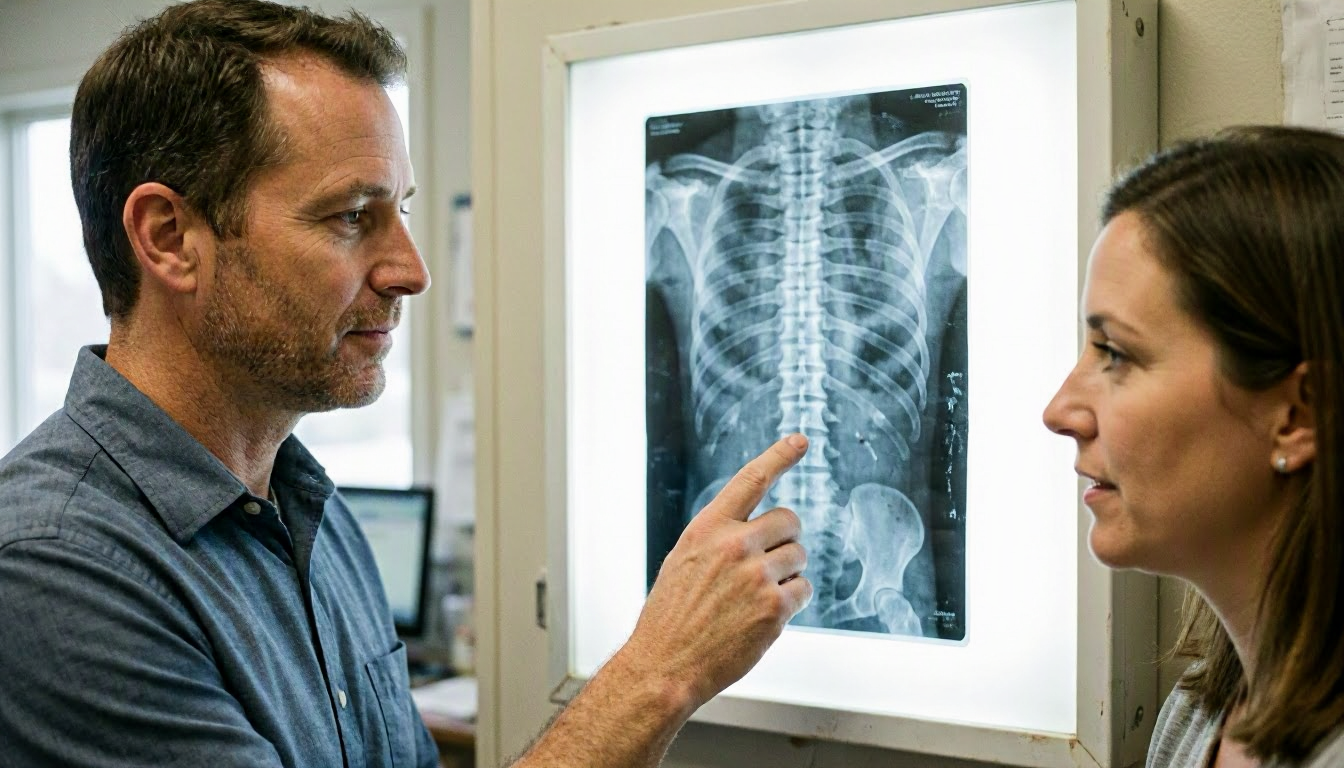
Chiropractic X-ray is a tool chiropractors use to see inside the body, especially the spine, bones. And joints. The images help chiropractors find problems like misalignments, fractures. Or arthritis that may cause pain or affect how the body moves. Unlike regular X-rays taken in hospitals, chiropractic X-rays focus on posture and spinal curves to guide treatment plans. They're usually quick, taking only a few minutes. And involve low levels of radiation.
Related glossary terms: Spinal Alignment, Chiropractic Adjustment, Cervical spine.
Chiropractors use X-rays to make sure adjustments are safe and effective. For example, if a patient has a history of trauma or severe pain, an X-ray can show if Common options include fractures or other issues that need special care. It also helps chiropractors see how the spine has changed over time, which can explain why someone might have chronic pain or limited movement. Not every patient needs an X-ray. But when they do, it provides important information that hands-on exams alone can't reveal.
How Chiropractic X-ray Works?
Chiropractic X-rays work by sending a small amount of radiation through the body. Dense structures like bones absorb more radiation and appear white on the image. While softer tissues appear darker. The chiropractor or X-ray technician positions the patient carefully to get clear pictures of specific areas, such as the neck, lower back. Or pelvis. Sometimes, multiple angles are needed to see the spine from the front, side. Or even a tilted view.
The images are reviewed right away to check for quality. If they are blurry or don't show the right area, the X-ray may need to be repeated. Once the images are clear, the chiropractor looks for signs of misalignment, degeneration. Or other issues. They may measure angles between bones or check the spacing between vertebrae to see if nerves could be compressed. This information helps them decide which adjustments or therapies will work best for the patient.
Why Chiropractic X-ray Matters?

Chiropractic X-rays matter because they help chiropractors make informed decisions about treatment. Without imaging, chiropractors rely only on physical exams, which can miss hidden problems like small fractures, bone spurs. Or severe arthritis. X-rays also help avoid risks by showing conditions that might make certain adjustments unsafe, such as osteoporosis or spinal instability. This makes treatment safer and more effective for patients.
For patients, X-rays can also provide clear next steps. Seeing the problem on an image helps them understand why they have pain or limited movement. In practical terms, it can also show progress over time, such as improved alignment after several adjustments. While X-rays are not needed for every patient, they are a valuable tool when there are questions about the cause of pain or the best way to treat it.
When Chiropractic X-ray Matters Most?
Chiropractic X-rays are most important when a patient has severe pain, a history of trauma. Or symptoms that do not improve with initial treatment. For example, if someone has been in a car accident or fallen, an X-ray can check for fractures or dislocations that need careful handling. They are also useful for patients with chronic conditions like scoliosis or arthritis, where the spine may have changed shape over time. In these cases, X-rays help chiropractors tailor treatment to avoid making the problem worse.
X-rays are also helpful for patients who have not responded to other treatments. If someone has tried physical therapy or medication without relief, an X-ray might reveal an underlying issue like a herniated disc or bone spur that needs a different approach. And chiropractors may use X-rays to monitor progress in patients with long-term spinal problems, ensuring that adjustments are helping and not causing harm. In Nashville, where many patients seek chiropractic care for work injuries or auto accidents, X-rays play a key role in accurate diagnosis and treatment planning.